Structure
The anatomy of muscles includes gross anatomy, which comprises all the muscles of an organism, and microanatomy, which comprises the structures of a single muscle.
Types
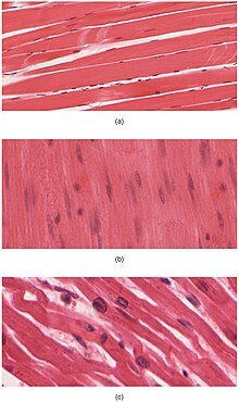
Muscle tissue is a soft tissue, and is one of the four fundamental types of tissue present in animals. There are three types of muscle tissue recognized in vertebrates:
- Skeletal muscle or "voluntary muscle" is anchored by tendons (or by aponeuroses at a few places) to bone and is used to effect skeletal movement such as locomotion and in maintaining posture. Though this postural control is generally maintained as an unconscious reflex, the muscles responsible react to conscious control like non-postural muscles. An average adult male is made up of 42% of skeletal muscle and an average adult female is made up of 36% (as a percentage of body mass).
- Smooth muscle or "involuntary muscle" is found within the walls of organs and structures such as the esophagus, stomach, intestines, bronchi, uterus, urethra, bladder, blood vessels, and the arrector pili in the skin (in which it controls erection of body hair). Unlike skeletal muscle, smooth muscle is not under conscious control.
- Cardiac muscle (myocardium), is also an "involuntary muscle" but is more akin in structure to skeletal muscle, and is found only in the heart.
Cardiac and skeletal muscles are "striated" in that they contain sarcomeres that are packed into highly regular arrangements of bundles; the myofibrils of smooth muscle cells are not arranged in sarcomeres and so are not striated. While the sarcomeres in skeletal muscles are arranged in regular, parallel bundles, cardiac muscle sarcomeres connect at branching, irregular angles (called intercalated discs). Striated muscle contracts and relaxes in short, intense bursts, whereas smooth muscle sustains longer or even near-permanent contractions.
Skeletal Muscle Fiber Types
The muscle fibers embedded in skeletal muscle are relatively classified into a spectrum of types given their morphological and physiological properties. Given a certain assortment of these properties, muscle fibers are categorized as slow-twitch (low force, slowly fatiguing fibers), fast twitch (high force, rapidly fatiguing fibers), or somewhere in between those two types (i.e. intermediate fibers). Some of the defining morphological and physiological properties used for the categorization of muscle fibers include: the number of mitochondria contained in the fiber, the amount of glycolytic, lipolytic, and other cellular respiration enzymes, M and Z band characteristics, energy source (i.e. glycogen or fat), histology color, and contraction speed and duration. Note that there is no standard procedure for classifying muscle fiber types. The properties chosen for classification depends on the particular muscle. For example, the properties used for distinguishing fast, intermediate, and slow muscle fibers can be different for invertebrate flight and jump muscle. To further complicate this classification scheme, the mitochondria content and other morphological properties within a muscle fiber can change with exercise and age.
Vertebrate Skeletal Muscle Fiber Types
- Type I, slow twitch, or "red" muscle, is dense with capillaries and is rich in mitochondria and myoglobin, giving the muscle tissue its characteristic red color. It can carry more oxygen and sustain aerobic activity using fats or carbohydrates as fuel. Slow twitch fibers contract for long periods of time but with little force.
- Type II, fast twitch muscle, has three major subtypes (IIa, IIx, and IIb) that vary in both contractile speed and force generated. Fast twitch fibers contract quickly and powerfully but fatigue very rapidly, sustaining only short, anaerobic bursts of activity before muscle contraction becomes painful. They contribute most to muscle strength and have greater potential for increase in mass. Type IIb is anaerobic, glycolytic, "white" muscle that is least dense in mitochondria and myoglobin. In small animals (e.g., rodents) this is the major fast muscle type, explaining the pale color of their flesh.
The density of mammalian skeletal muscle tissue is about 1.06 kg/liter. This can be contrasted with the density of adipose tissue (fat), which is 0.9196 kg/liter. This makes muscle tissue approximately 15% denser than fat tissue.
Microanatomy
Skeletal muscles are sheathed by a tough layer of connective tissue called the epimysium. The epimysium anchors muscle tissue to tendons at each end, where the epimysium becomes thicker and collagenous. It also protects muscles from friction against other muscles and bones. Within the epimysium are multiple bundles called fascicles, each of which contains 10 to 100 or more muscle fibers collectively sheathed by a perimysium. Besides surrounding each fascicle, the perimysium is a pathway for nerves and the flow of blood within the muscle. The threadlike muscle fibers are the individual muscle cells (myocytes), and each cell is encased within its own endomysium of collagen fibers. Thus, the overall muscle consists of fibers (cells) that are bundled into fascicles, which are themselves grouped together to form muscles. At each level of bundling, a collagenous membrane surrounds the bundle, and these membranes support muscle function both by resisting passive stretching of the tissue and by distributing forces applied to the muscle. Scattered throughout the muscles are muscle spindles that provide sensory feedback information to the central nervous system. (This grouping structure is analogous to the organization of nerves which uses epineurium, perineurium, and endoneurium).
This same bundles-within-bundles structure is replicated within the muscle cells. Within the cells of the muscle are myofibrils, which themselves are bundles of protein filaments. The term "myofibril" should not be confused with "myofiber", which is a simply another name for a muscle cell. Myofibrils are complex strands of several kinds of protein filaments organized together into repeating units called sarcomeres. The striated appearance of both skeletal and cardiac muscle results from the regular pattern of sarcomeres within their cells. Although both of these types of muscle contain sarcomeres, the fibers in cardiac muscle are typically branched to form a network. Cardiac muscle fibers are interconnected by intercalated discs, giving that tissue the appearance of a syncytium.
The filaments in a sarcomere are composed of actin and myosin.
Gross anatomy
The gross anatomy of a muscle is the most important indicator of its role in the body. There is an important distinction seen between pennate muscles and other muscles. In most muscles, all the fibers are oriented in the same direction, running in a line from the origin to the insertion. However, In pennate muscles, the individual fibers are oriented at an angle relative to the line of action, attaching to the origin and insertion tendons at each end. Because the contracting fibers are pulling at an angle to the overall action of the muscle, the change in length is smaller, but this same orientation allows for more fibers (thus more force) in a muscle of a given size. Pennate muscles are usually found where their length change is less important than maximum force, such as the rectus femoris.
Skeletal muscle is arranged in discrete muscles, an example of which is the biceps brachii (biceps). The tough, fibrous epimysium of skeletal muscle is both connected to and continuous with the tendons. In turn, the tendons connect to the periosteum layer surrounding the bones, permitting the transfer of force from the muscles to the skeleton. Together, these fibrous layers, along with tendons and ligaments, constitute the deep fascia of the body.
Muscular system
The muscular system consists of all the muscles present in a single body. There are approximately 650 skeletal muscles in the human body, but an exact number is difficult to define. The difficulty lies partly in the fact that different sources group the muscles differently and partly in that some muscles, such as palmaris longus, are not always present.
A muscular slip is a narrow length of muscle that acts to augment a larger muscle or muscles.
The muscular system is one component of the musculoskeletal system, which includes not only the muscles but also the bones, joints, tendons, and other structures that permit movement.
Development
All muscles are derived from paraxial mesoderm. The paraxial mesoderm is divided along the embryo's length into somites, corresponding to the segmentation of the body (most obviously seen in the vertebral column. Each somite has 3 divisions, sclerotome (which forms vertebrae), dermatome (which forms skin), and myotome (which forms muscle). The myotome is divided into two sections, the epimere and hypomere, which form epaxial and hypaxial muscles, respectively. The only epaxial muscles in humans are the erector spinae and small intervertebral muscles, and are innervated by the dorsal rami of the spinal nerves. All other muscles, including those of the limbs are hypaxial, and inervated by the ventral rami of the spinal nerves.
During development, myoblasts (muscle progenitor cells) either remain in the somite to form muscles associated with the vertebral column or migrate out into the body to form all other muscles. Myoblast migration is preceded by the formation of connective tissue frameworks, usually formed from the somatic lateral plate mesoderm. Myoblasts follow chemical signals to the appropriate locations, where they fuse into elongate skeletal muscle cells.


Comments
Post a Comment